Newly created software provides diagnostic support to radiologists and other professionals engaged in CT scan interpretation. This software was developed utilizing deep learning, a form of artificial intelligence (AI).
This innovation involves software that has been trained to translate interpretations from MRI images into corresponding CT images of brain cases.
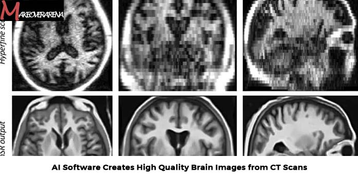
AI Software Creates High Quality Brain Images from CT Scans
Computed tomography (CT) is a cost-effective imaging technology accessible in healthcare facilities, including regions with limited access to other imaging methods. However, it is often seen as less capable than magnetic resonance imaging (MRI) in visualizing complex brain structural changes and variations in ventricular system flow. As a result, certain imaging tasks necessitate specialized units within larger hospitals that possess advanced imaging technology.
Dependable Decision Making
Michael Schöll, a professor at Sahlgrenska Academy who led the study in collaboration with researchers from Karolinska Institutet, the National University of Singapore, and Lund University, expressed:
- Our approach produces diagnostically valuable information from standard CT scans, which, in certain instances, rivals the quality of an MRI scan conducted in specialized healthcare settings.
- The key idea is that this straightforward and rapid method can yield a wealth of information from routine examinations performed in primary care as well as specific investigations within specialist healthcare.
- In its early phase, this method can aid in dementia diagnosis, but it is also expected to find applications in other areas of neuroradiology.
According to the researchers, this marks a clinically proven use of AI-driven algorithms that could evolve into a swift and reliable decision support tool, reducing the chances of false negatives.
As a result, these researchers expect that this solution could improve diagnostic capabilities in primary care, ultimately making patient referrals to specialist care more efficient.
- This signifies a significant advancement in imaging diagnosis,” remarked Meera Srikrishna, a postdoctoral fellow at the University of Gothenburg and the lead author of the study, published in the journal Alzheimer’s & Dementia.
- Now, it is feasible to measure the dimensions of various brain structures or regions in a manner similar to the advanced analysis of MRI images. The software enables the segmentation of the brain’s components in the image and the measurement of its volume, even when the image quality is not on par with that of CT scans.
The software was created by utilizing images from a group of 1,117 individuals, all of whom had undergone both CT and MRI scans. The primary emphasis of the current study revolved around healthy older adults and patients with various forms of dementia.


